Virus and Virus-Like Particle
Viruses and virus-like particles (VLPs) play a crucial role in therapeutic and diagnostic innovations, offering immense potential for vaccine development, advanced therapies, and groundbreaking research. Particle Metrix’s cutting-edge Nanoparticle Tracking Analysis (NTA) systems provide precise, reliable, and comprehensive characterization of these particles, including size, concentration, and fluorescence-based analysis — all within a single measurement.
With unparalleled accuracy and advanced features like colocalization, our systems empower researchers to drive critical breakthroughs in virology and pharmaceutical development, making Particle Metrix the ideal partner for virus and VLP analysis.
Understanding Viruses and Virus-Like Particles
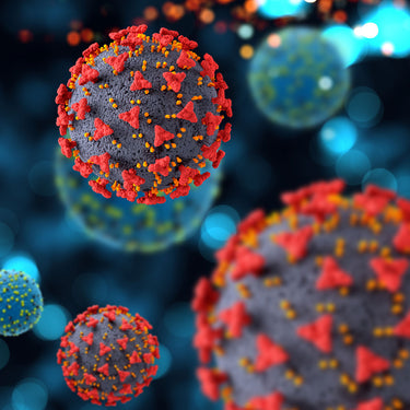
Viruses are nanoscale pathogens capable of infecting humans, animals, plants, and bacteria. Lacking independent metabolism, they rely entirely on host cells for replication. They exist either within host cells or as independent virions, consisting of genetic material encased in a protein shell called a capsid. Some viruses, such as SARS-CoV-2 and HIV, also have a lipid bilayer envelope derived from their host cells.
VLPs are engineered structures designed to mimic viral architecture while lacking genetic material, rendering them non-infectious. This makes them particularly suitable for safe therapeutic applications like vaccines. Their sizes vary:
- Influenza virus: 80–120 nm
- SARS-CoV-2: 60–140 nm
- Small viruses (e.g., Circoviridae): 15–30 nm
- Large viruses (e.g., giant viruses): 400–500 nm
Both viruses and VLPs are integral to virology research, therapeutics, and vaccine development.

Types of Viruses and Virus-Like Particles
Viruses and VLPs can be categorized based on their structure and intended use:
Enveloped types: Contain a lipid bilayer, such as influenza and SARS-CoV-2.
Non-enveloped types: Composed solely of protein shells, like adenoviruses and HPV.
Hybrid VLPs: Custom-engineered for applications in drug delivery and gene therapy.

Challenges in research and characterization
The study of viruses and VLPs presents unique challenges due to their small size, structural complexity, and heterogeneity. Isolation and characterization are often difficult due to contaminants like proteins, lipoproteins, and cellular debris. Additionally, their tendency to aggregate complicates accurate measurement and may impact functionality.
Identifying subpopulations within viral particle samples is critical for both research and therapeutic applications. Fluorescence-based techniques such as Nanoparticle Tracking Analysis (F-NTA) enable precise detection of specific viral markers, enhancing analytical accuracy.

Advanced Analysis with NTA
Sophisticated analytical tools are essential for measuring size, concentration, and stability of viral particles with high precision. NTA is a gold-standard technique that provides unparalleled insights into their behavior and properties.
Our ZetaView® PMX-230 TWIN and PMX-420 QUATT systems offer high-resolution measurements of particle size, concentration, and zeta potential. Fluorescence detection and colocalization capabilities enable accurate quantification of subpopulations by tagging viral markers such as capsid proteins or envelope glycoproteins.
ZetaView® Analyzer
ZetaView®for Virus and VLP Analysis
The ZetaView® systems from Particle Metrix are designed to address the unique challenges of virus and VLP research. Key features include:
High-Resolution Measurement
Provides precise data on size, concentration, and zeta potential.
Fluorescence Capabilities
Enables identification and quantification of subpopulations with specificity.
Colocalization Analysis
Allows researchers to detect multiple markers on individual particles for in-depth sample characterization.

Therapeutic and Research Applications
Vaccine Development:
VLPs serve as safe and effective platforms for vaccines against diseases such as hepatitis B, HPV, and COVID-19, eliciting robust immune responses without infection risk.
Gene Therapy:
Viral vectors enable precise delivery of genetic material to treat hereditary diseases.
Oncolytic Cancer Therapy:
Certain viruses are designed to selectively target and destroy cancer cells while sparing healthy tissue.

Industries utilizing Virus and VLP Technology
Biomedical Research
Essential for studying infection mechanisms, immune responses, and disease progression, leading to advancements in antiviral therapies, vaccines, and diagnostics.
Pharmaceutical Industry
Critical for vaccine development, gene therapy, and biologics, ensuring product safety and efficacy.
Biotechnology
Used in personalized medicine, oncolytic cancer therapies, and innovative drug delivery systems.
Diagnostic Laboratories
Support the development of precise testing methods for infectious diseases and aid in monitoring treatment efficacy and viral mutations.
Healthcare
Play a key role in advancing treatments and improving patient outcomes, from vaccines to targeted therapies.
Academic Research Institutions
Drive fundamental discoveries in virology and translate basic research into clinical applications.
Unlock the full potential of your Virology Research
Viruses and VLPs are at the forefront of modern science, with applications spanning diagnostics, therapeutics, and beyond. Their analysis requires precision and reliability—qualities that Particle Metrix’s advanced NTA systems deliver.
Take your virus and VLP research to the next level with Particle Metrix. Contact us today to schedule a consultation or live demo.
Frequently asked questions
What is the typical size of viruses like the flu or SARS-CoV-2?
- Influenza virus: 80–120 nm
- SARS-CoV-2: 60–140 nm
How are viruses and VLPs characterized?
Characterization involves measuring size, concentration, and subpopulations using advanced techniques such as fluorescence-based NTA and colocalization.
What are the medical applications of viruses and VLPs?
Viruses and VLPs are used in vaccines, gene therapy, oncolytic cancer treatments, and phage therapy due to their structural versatility and safety profiles.