Extracellular Vesicles analysis
Extracellular Vesicles (EVs) are increasingly being recognized as key players in cellular communication and disease research, offering immense potential in diagnostics and therapeutics. Particle Metrix’s advanced Nanoparticle Tracking Analysis systems are specifically designed to deliver precise, reliable, and detailed analyses of Extracellular Vesicles, including size, concentration, and fluorescence-based phenotyping, all within a single measurement.
With unmatched accuracy and innovative features like colocalization, our systems empower researchers to make critical breakthroughs in biomedical and pharmaceutical applications, making them the ideal choice for Extracellular Vesicle analysis.
What are Extracellular Vesicles?
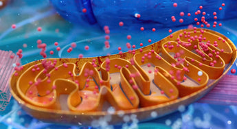
Extracellular Vesicles are small, lipid-bilayer nanoparticles released by both eukaryotic and prokaryotic cells, with sizes ranging from 30 nm to 4 µm. First identified in 1956 by Don Fawcett during electron microscopy of tumor cells, their importance was not fully realized until recent decades due to the lack of appropriate analysis techniques. The development of Nanoparticle Tracking Analysis marked a turning point, as it allowed Extracellular Vesicles to be visualized in their natural, dispersed state, significantly advancing our understanding of their biological functions.
Nanoparticle Tracking Analysis and other methods have shown that Extracellular Vesicles are essential in intercellular communication, transporting DNA, RNA, proteins, and lipids between cells. These vesicles are now recognized for their critical roles in regulating cellular processes and are a key focus in biomedical research, particularly for their potential as diagnostic biomarkers and therapeutic agents in various diseases.
Types of Extracellular Vesicles
Extracellular Vesicles are categorized into three main types based on their size and formation:
Exosomes
Ranging from 30–130 nm, exosomes are of endosomal origin and released via exocytosis.
Microvesicles
Formed through outward budding of the plasma membrane, these vesicles can be as large as 1,000 nm.
Apoptotic Bodies
The largest of the Extracellular Vesicles, reaching up to 4 µm, formed during programmed cell death.
Challenges in Extracellular Vesicles research and characterization
While Extracellular Vesicles offer significant potential for diagnostics and therapeutics, their characterization poses technical challenges. Extracellular Vesicles are difficult to isolate due to their small size and the presence of contaminating particles like lipoproteins.
Accurate and reproducible characterization requires advanced techniques like Nanoparticle Tracking Analysis and fluorescence-based Nanoparticle Tracking Analysis (F-NTA), which allow for the identification of specific Extracellular Vesicles subpopulations in complex biological samples. Fluorescence colocalization (C-NTA) further enhances this by detecting multiple markers on the same Extracellular Vesicles.
Analyzing Extracellular Vesicles
The analysis of Extracellular Vesicles requires precise and advanced techniques to measure their size, concentration and subpopulation distribution. Nanoparticle Tracking Analysis has become a leading method for studying Extracellular Vesicles, delivering critical insights into their behavior in biological systems. Our innovative ZetaView® systems, provides enhanced capabilities tailored to the challenges of Extracellular Vesicles analysis.
By combining high-resolution size and concentration measurements with advanced zeta potential and fluorescence detection, ZetaView® systems provide researchers with a comprehensive toolkit for understanding Extracellular Vesicles. These systems represent the cutting edge of Extracellular Vesicles analysis, empowering researchers to explore their role in biological systems with unmatched precision and detail.
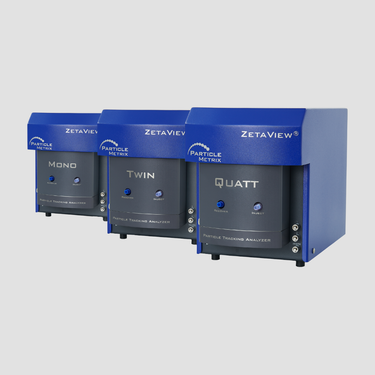
ZetaView® Analyzer
ZetaView® for Extracellular Vesicles analysis
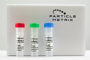
The ZetaView® PMX-230 TWIN and PMX-420 QUATT systems are specifically optimized for high-precision measurements of Extracellular Vesicles size, concentration and zeta potential. These advanced Nanoparticle Tracking Analysis solutions integrate dual- or quadruple-laser fluorescence detection, enabling researchers to perform detailed phenotyping and accurately quantify Extracellular Vesicles subpopulations by labeling specific markers such as CD9 and CD63.
The colocalization capabilities further enhance specificity, offering unparalleled insights into Extracellular Vesicles subpopulation behavior.
Which function do Extracelluar Vesicles have?
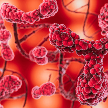
Extracellular Vesicles are critical in facilitating intercellular communication, playing diverse roles across biological systems. Their ability to transfer molecular cargo, including proteins, lipids, and nucleic acids in extracellular spaces is assumed to influence a wide range of cellular processes. The field of EV research is still relatively new and the depth of the significance of Extracellular Vesicles has not yet been explored. But it is already clear that EVs are promising candidates for a multitude of diagnostic and therapeutic applications.
Immune Modulation
Extracellular Vesicles play a pivotal role in modulating immune responses by acting as carriers of antigens or immune regulatory molecules. They can present antigens to immune cells, thus initiating or regulating immune responses. This function is especially important in contexts such as infections, where Extracellular Vesicles can either enhance the body’s defense mechanisms or, in some cases, be co-opted by pathogens to evade immune detection and facilitate infection. In autoimmune diseases, Extracellular Vesicles may contribute to immune dysregulation by spreading inflammatory signals.
Disease Mechanisms
Extracellular Vesicles are heavily involved in the pathogenesis of several major diseases, making them prime targets for both diagnostics and therapeutics. In cancer, tumor-derived Extracellular Vesicles (also known as exosomes) facilitate tumor growth, metastasis, and resistance to therapies by transferring oncogenic signals to surrounding cells.
These Extracellular Vesicles can also modulate the immune environment around tumors, making it more favorable for cancer progression. Furthermore, cancer-derived Extracellular Vesicles contribute to drug resistance, enabling tumor cells to evade conventional treatments.
In cardiovascular diseases, Extracellular Vesicles play dual roles, contributing to both protective and harmful processes. They can mediate inflammation and promote the formation of blood clots (thrombosis) in conditions like atherosclerosis and stroke, thereby exacerbating disease progression. At the same time, certain types of Extracellular Vesicles have been found to aid in repairing damaged blood vessels, offering protective benefits in some cardiovascular contexts.
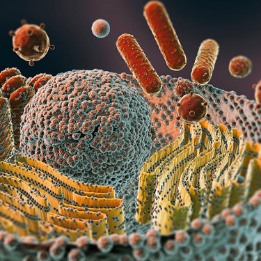
In infectious diseases, Extracellular Vesicles are involved in both host defense and pathogen propagation. Host-derived Extracellular Vesicles play a role in modulating the immune response, helping to fight infections by transporting immune signals. However, pathogen-derived Extracellular Vesicles can manipulate host immune responses and promote disease progression by transferring virulence factors between host cells, thereby enhancing the infection's spread. This dual functionality highlights the complexity of Extracellular Vesicles' role in infectious disease dynamics.
By understanding these mechanisms, researchers are exploring Extracellular Vesicles as both diagnostic biomarkers and drug-delivery platforms, particularly in cancer, cardiovascular, and infectious diseases.
Tissue Regeneration
One of the most promising therapeutic applications of Extracellular Vesicles is in tissue repair. Extracellular Vesicles derived from stem cells, particularly mesenchymal stem cells (MSCs), carry growth factors and other regenerative molecules that promote healing and tissue regeneration. These Extracellular Vesicles can enhance the repair of damaged tissues, such as in wound healing or recovery after cardiac injury.

Industries where Extracellular Vesicles are relevant
Extracellular Vesicles research is critical across several industries due to the versatile nature of Extracellular Vesicles and their potential role in diagnostics and therapeutics:
Biomedical Research
Extracellular Vesicles are key to understanding disease mechanisms and developing novel treatments.
Pharmaceutical Industry
Extracellular Vesicles are being explored as vehicles for drug delivery, due to their ability to transfer molecular cargo between cells.
Diagnostic Laboratories
Extracellular Vesicles serve as biomarkers for diseases, including cancer and cardiovascular conditions, offering potential for early diagnosis.
Universities and Academic Research Institutes
Extracellular Vesicles are a primary focus in basic and applied research, contributing to the understanding of cellular processes and disease progression.
Let’s talk about your Extracellular Vesicles Analysis needs
Extracellular Vesicles are central to both fundamental research and applied sciences, ranging from diagnostics to therapeutic development. With applications in biomedical research, pharmaceutical development and diagnostics, we are the ideal partner for labs aiming to excel in Extracellular Vesicles analysis.
Unlock the full potential of your Extracellular Vesicles research with Particle Metrix. Contact us today to schedule a free consultation or live demo.
Frequently asked questions about Extracellular Vesicles
Are exosomes Extracellular Vesicles?
Yes, exosomes are a specific type of extracellular vesicle. They are the smallest extracellular vesicles, typically ranging from 30 to 130 nm in size, and are formed within the endosomal system of cells. Exosomes are released into the extracellular environment through a process called exocytosis, where they play key roles in cell-to-cell communication and the transport of molecular cargo.
How are Extracellular Vesicles formed?
Extracellular Vesicles are formed through different mechanisms depending on their type. Exosomes are generated within multivesicular bodies in the endosomal system and released when these bodies fuse with the cell membrane. Microvesicles, on the other hand, form by directly budding off from the plasma membrane. Apoptotic bodies are released during programmed cell death (apoptosis) as the cell breaks down. Each type of Extracellular Vesicle carries molecular signals that can affect target cells in various ways.
How to isolate Extracellular Vesicles?
Isolating Extracellular Vesicles typically involves separating them from other cellular components using techniques such as ultracentrifugation, size exclusion chromatography, and precipitation methods. Each method offers different advantages depending on the sample and required purity. However, after isolation, Nanoparticle Tracking Analysis (NTA) is widely considered the easiest and fastest technology for verifying and characterizing extracellular vesicles. NTA is one of the main technologies recommend by the MISEV guidelines.